Toward real world medical image analysis
Comprehensive Analysis & computing of REal-world medical images (CARE2026)
CARE 2026 has successfully concluded. Thank you all for your active participation! By 2026.08.26, we have 279 teams participated, and received 48 paper submission. You can refer to our General Info Page for more details. Leaderboards will be released after the workshop.
Contact: If you have any questions, please contact care26challenge@163.com or care2026challenge@outlook.com.
CARE 2026 accepted papers (29)
(NEW!) CARE 2026 Workshop Program
- 📍 Room: Strasbourg Convention Center, Room Auditorium Cassin (G) , Strasbourg, France
- 📅 Date: Sunday, 27 September 2026
- 🕒 Time: 15:30 – 18:00
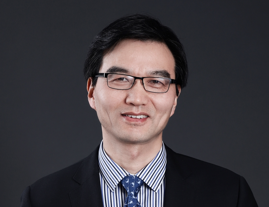
Prof. Dinggang Shen
School of BME, ShanghaiTech University, China
Shanghai United Imaging Intelligence Co., Ltd., China
Dinggang Shen is a Professor and a Founding Dean with School of Biomedical Engineering, ShanghaiTech University, and also a Co-CEO of United Imaging Intelligence (UII), Shanghai, China. He is a Fellow of IEEE, AIMBE, IAPR, MICCAI, ISMRM, and IAMBE. He was a recipient of the Distinguished Investigator Award from The Academy for Radiological & Biomedical Imaging Research, USA (2019), Technical Achievement Award from IEEE EMBS (2024), and Enduring Impact Award from MICCAI (2025). He was a Jeffrey Houpt Distinguished Investigator and a Full Professor (Tenured) with University of North Carolina at Chapel Hill, USA. He was also a tenure-track assistant professor in University of Pennsylvanian, and an Instructor in Johns Hopkins University. His research interests include medical image analysis and artificial intelligence, in which he published >800 SCI papers, with H-index 172 and 120K+ citations. He serves as Senior Editor for Medical Image Analysis, IEEE TBME, and IEEE TMI, and also editorial board member for six international journals. He has served in the Board of Directors, The Medical Image Computing and Computer Assisted Intervention (MICCAI) Society, in 2012-2015, and was the General Chair for MICCAI 2019.
“Development and Deployment of Multimodal AI Foundation Models for Real-World Clinical Workflows”
Abstract: Conventional deep learning methods have been extensively investigated throughout end-to-end clinical workflows to support medical imaging, disease screening, clinical diagnosis, treatment planning, and prognostic prediction. Nevertheless, such task-specific models require separate development and optimization for individual clinical scenarios, leading to inefficient model development. Multimodal AI foundation models have fundamentally shifted this research paradigm by enabling a single unified model to accommodate multiple heterogeneous clinical tasks, greatly expediting AI development and real-world clinical translation. This talk will review traditional deep learning applications in routine clinical practice and present our recent progress in foundation model-enabled medical imaging solutions, demonstrating their promising capability to enhance clinical workflows.
Posters
Note: The poster should be in PORTRAIT format and not exceed A0 size (33.1 in x 46.8 in or 841 mm x 1189 mm).
Motivation
Medical image analysis empowered by AI has significantly reshaped medicine. However, real-world medical image analysis poses significant challenges for medical AI, including domain shifts due to diverse imaging protocols and medical centers, variability in pathological regions of interest (ROIs) such as lesions and scars, and limited generalization capabilities of existing deep learning models. This challenge aims to encourage the development of novel and effective methods that facilitate the translation of medical AI into real-world clinical practice.
Tracks
This challenge consists of 4 challenging tracks and encompasses imaging data from over 2000 patients collected across three continents, capturing diverse acquisition protocols, clinical centers, and patient populations. Participants can explore each track below to access the corresponding datasets and detailed task descriptions.
CARE-Left Atrium: Left Atrial Segmentation and Analysis.
CARE-Liver: Liver Segmentation and Fibrosis Staging.
CARE-Myocardium: Myocardial Pathology Segmentation.
CARE-Whole Heart: Whole Heart Segmentation.
Submission
After registration, we will assign participants an account to login into our evaluation platform. Participants can directly upload your predictions and docker via the website. For paper submission, please refer to our paper submission guidance.
Post-Challenge Data Request
For post-challenge data access requests, please contact care26challenge@163.com.
Timeline
The schedule for this track is as follows. All deadlines (DDLs) are on 23:59 in Pacific Standard Time.
| Registration Period | March 27, 2026 - July 20, 2026 |
|---|---|
| Training Data Release | March 27, 2026 |
| Validation Phase start | May 01, 2026 |
| Test data release | June 10, 2026 |
| Test Results Submission | August 03, 2026 (Updated!) |
| Abstract Submission | July 27, 2026 (Updated!) |
| Paper Submission | July 27, 2026 (Updated!) |
| Paper Acceptance Notification | August 10, 2026 |
| Camera Ready | August 24, 2026 |
| Workshop (Half-Day) | September 27, 2026 |
Awards
The challenge has 1 best paper and 2 runner-ups. In addition, CARE 2026 will provide 8 best performance awards as follows:
CARE-Left Atrium:
- LA scar quantification (MRI)
- LA multi-structure segmentation (CT)
CARE-Liver:
- Liver Fibrosis Staging (LiFS)
- Liver Segmentation (LiSeg)
CARE-Myocardium:
- Myocardial Pathology Segmentation
- Cine Myocardial Pathology Segmentation
CARE-Whole Heart:
- Whole heart segmentation (CT)
- Whole heart segmentation (MRI)
Sponsors
- MICCAI-SIG Cardiac Imaging, Computational Modelling and Clinical Sciences (SIG-Cardiac)
- MICCAI-SIG Explainable AI for Medical Image Analysis (SIG-xMedIA)